Le tendinopatie croniche e acute sono difficili da trattare e la guarigione dei tendini è generalmente un processo molto lento e incompleto e la nostra comprensione generale della biologia dei tendini e della rigenerazione è in ritardo rispetto a quella dei muscoli o delle ossa. Sebbene ancora in gran parte inesplorati, diversi studi suggeriscono un effetto positivo degli interventi nutrizionali sulla salute e riparazione dei tendini. Con questo studio, miriamo a rivelare gli effetti di una dieta ricca di glucosio sulla neoformazione del tendine in un modello di ratto non diabetico di tenotomia di Achille. Dopo l'intervento chirurgico, gli animali hanno ricevuto una dieta ricca di glucosio o una dieta di controllo rispettivamente per 2 e 4 settimane. Rispetto al gruppo di controllo, lo spessore e la rigidità del tessuto di riparazione del tendine erano aumentati nel gruppo ad alto contenuto di glucosio dopo 2 settimane e il modello dell'andatura era alterato dopo 1 e 2 settimane. La proliferazione cellulare era fino a 3 volte superiore e l'espressione dei geni marcatori condrogenici Sox9, Col2a1, Acan e Comp era significativamente aumentata 2 e 4 settimane dopo l'intervento chirurgico. Inoltre, dopo 4 settimane di una dieta ricca di glucosio era evidente un moderato aumento delle aree simili alla cartilagine all'interno del tessuto di riparazione. In sintesi, proponiamo che una dieta ricca di glucosio influenzi significativamente la guarigione dei tendini dopo un infortunio nei ratti non diabetici, guidando potenzialmente la degenerazione condrogena.
Stefanie Korntner 1 2,
Nadja Kunkel 1 2 3,
Christine Lehner 1 2,
Renate Gehwolf 1 2,
Andrea Wagner 1 2,
Pietro Augusto 4,
Daniele Stefano 4,
Verena Heu 5,
Hans Christian Bauer 1 2,
Andrea Traweger 1 2,
Herbert Tempfer 6 7 Affiliazioni
- 1Institute of Tendon & Bone Regeneration, Paracelsus Medical University di Salisburgo, Centro per le lesioni del midollo spinale e la rigenerazione dei tessuti di Salisburgo, Salisburgo, AT, Austria.
- 2Cluster austriaco per la rigenerazione dei tessuti, Vienna, AT, Austria.
- 3Ospedale Universitario di Salisburgo, Dipartimento di Chirurgia Traumatologia e Lesioni Sportive, Salisburgo, AT, Austria.
- 4Istituto di Biomeccanica, Trauma Center Murnau, Murnau, DE, Germania.
- 5Ospedale Universitario di Salisburgo, Dipartimento di Pediatria, Salisburgo, AT, Austria.
- 6Institute of Tendon & Bone Regeneration, Paracelsus Medical University di Salisburgo, Centro per le lesioni del midollo spinale e la rigenerazione dei tessuti di Salisburgo, Salisburgo, AT, Austria. herbert.tempfer@pmu.ac.at.
- 7Cluster austriaco per la rigenerazione dei tessuti, Vienna, AT, Austria. herbert.tempfer@pmu.ac.at.
Figure 1 Gait Analysis: Intermediate toe spread (ITS). Intermediate toe spread (ITS) of the right hind limb (RH) before surgery was equal between the experimental groups (
a). One week post-op, ITS was significantly higher in the high-glucose group compared to the control group (p < 0.0001; Mann-Whitney test, two-tailed). 2 weeks post-op, ITS remained significantly higher in the high-glucose group compared to the control diet group (p < 0.0001; Mann-Whitney test, two-tailed). Representative images for ITS measurements pre-op (
b, one week post-op (
c) and 2 weeks post-op (
d). ITS (thick white bar) represents the distance between second and fourth toe.
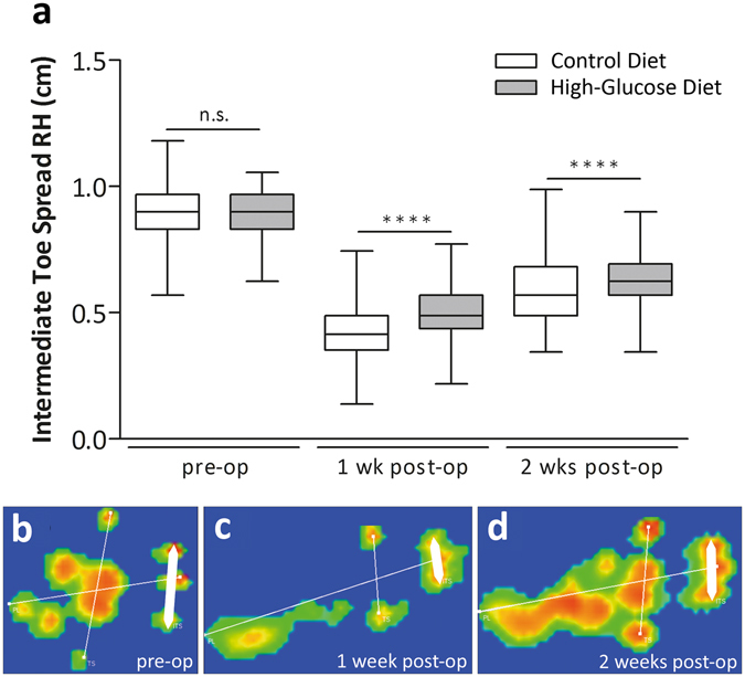 Figure 2
 Repair tissue thickness measurements. Comparisons among the experimental groups 2 weeks after surgery revealed significantly increased thickness in the high-glucose group (n = 25), compared to the control diet group (n = 24, p < 0.0001; Mann-Whitney test, two-tailed) (
a). 4 weeks after surgery no significant difference could be detected (n = 6; p = 0.0519; Mann-Whitney test, two-tailed). Tendon thickness was determined 3 mm proximal from the calcaneus (
b, black bars). Values for intact tendons are provided as a reference (n = 24).
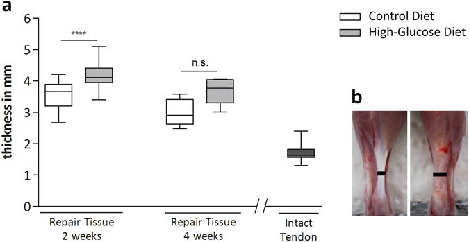 Figure 3
 Biomechanical properties of tendon repair tissues 2 weeks after Achilles tenotomy. Biomechanical tests comparing maximum tensile load of the repair tissues 2 weeks after surgery revealed no significant difference (p > 0.05; Mann-Whitney test, two-tailed) between the experimental groups (
a). Tendon stiffness measurements 2 weeks after surgery among the experimental groups showed significantly stiffer repair tissues (p < 0.05, asterisk; Mann-Whitney test, two-tailed) in the high-glucose group (n = 14) when compared to the control group (n = 14) (
b). Values for intact tendons are provided as a reference (n = 9). Representative force-elongation diagrams of the repair tissues of the control diet group (
c), the high-glucose diet group (
d) and of an intact tendon (
e). In the toe-region collagen fibres are crimped and become elongated and straightened with applied loading. The linear region represents tendon stiffness. Microscopic tears occur in the plastic region where the slope drops until the maximum tensile load is reached and the tendon ruptures.
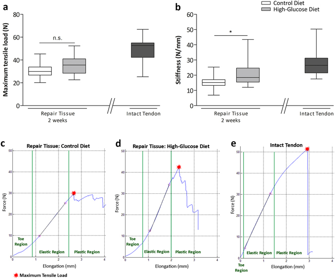 Figure 4
 Descriptive histology of tendon repair tissues 2 and 4 weeks post-surgery. Representative histologic images of tendon repair tissue sections of the control diet group and the high-glucose diet group stained with either Herovici’s polychrome (
a,
b,
e,
f), or Masson-Goldner trichrome (
c,
d,
g,
h). Loose collagen fibre orientation (c’), hypercellularity and ingrowth of vessels and nerves after 2 weeks (d’). The extracellular matrix (ECM) was mainly composed of immature collagen, most likely corresponding to collagen type III ((
a,
b,
e,
f); blue staining), both 2 and 4 weeks after surgery. Aggregates of cells with a chondrogenic phenotype (
arrows) were frequently observed 4 weeks post-surgery in both the control and the high-glucose diet group, although to a different extent (g’,h’). No difference could be detected between the experimental groups.
Arrows: retracted proximal and distal ends of the original tendon;
asterisks: newly formed tendon repair tissue.
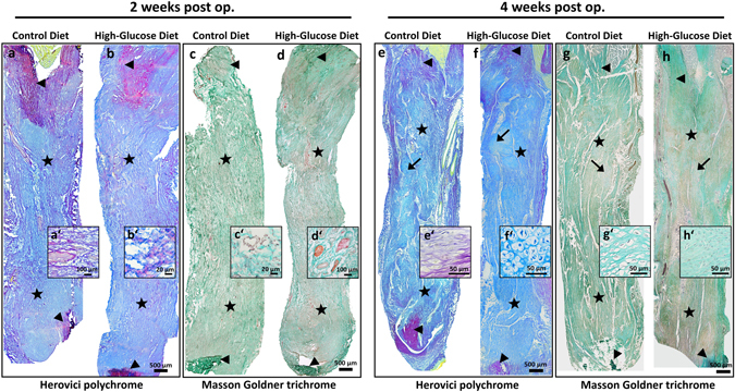 Figure 5
 Analysis of COL1 and COL3 and tenogenic marker gene expression. Quantitative PCR analysis of
Col1a1 (
a) and
Col3a1 (
b) and the tenogenic lineage associated genes
Mkx (
c),
Scx (
d), and
Tnmd (
e) in the repair tissues of the control diet group and the high-glucose diet group after 2 and 4 weeks. COL1 staining was mostly confined to capillaries within the repair tissue after 2 weeks whereas a deposition of collagen type 1 in the extracellular matrix of the repair tissue was observed after 4 weeks. Strong COL1 staining in intact tendon tissue is shown as a reference (
f). Substantial collagen type 3 deposition in the extracellular matrix was seen after 2 and 4 weeks. Note the strong COL3 staining in the epitenon (
asterisk) of intact tendon tissue (
g). Compared to intact tendons,
Tnmd expression was increased 2 and 4 weeks after surgery.
Mkx expression was lower in the repair tissues, showing no substantial change over time.
Scx expression 2 weeks post-tenotomy was comparable to intact tendons. 4 weeks post-surgery
Scx transcript levels were lower, the decrease being strongest for the high-glucose group. Values for intact tendons are provided as a reference (n = 12).
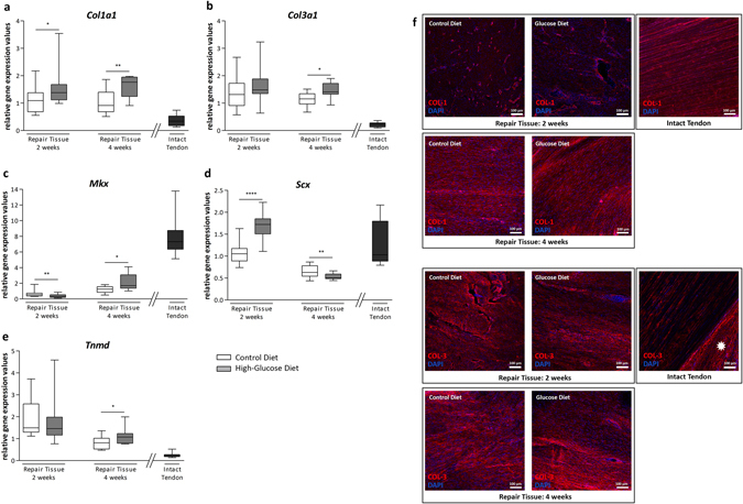 Figure 6
 Quantification of cell proliferation by KI67 in tendon repair tissues. Compared to a control diet group, a high-glucose diet significantly increases the proportion of KI67-positive cells after 2 (p < 0.0001, n = 15 images of 3 animals/group) and 4 weeks (p < 0.0001, n = 30 images of 3 animals/group) (
a) (Mann-Whitney test, two-tailed), whereas the total number of cells/area is significantly decreased in the high-glucose group compared to the control group after 2 weeks (
g) (p < 0.0001, Mann-Whitney test, two-tailed). Representative images for intact control tendons (
b) and tendon repair tissues 2 weeks post op: control group (
c), high-glucose group (
d) and 4 weeks post op: control group (
e) and high-glucose group (
f). Values for intact tendons are provided as a reference (n = 5 images of 3 animals).
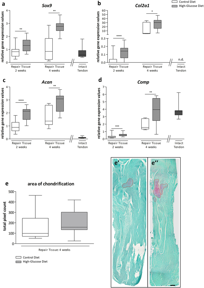
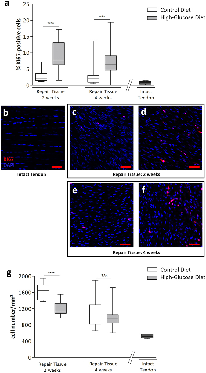 Figure 7
 Gene expression analysis and histomorphometry of tendon repair tissues. Quantitative PCR analysis of tendon repair tissues shows that the chondrocyte lineage-associated genes
Sox9 (
a),
Col2a1 (
b),
Acan (
c) and
Comp (
d) are significantly upregulated in animals receiving a high-glucose diet for 2 and 4 weeks, respectively, compared to animals fed a control diet (Mann-Whitney test, two-tailed). Values for intact tendons are provided as a reference. Quantification of areas showing beginning chondrification 4 weeks after surgery did show a trend of increasing chondrification in the high-glucose group (
e), though the difference was not significant (p = 0,328; Mann-Whitney test, two-tailed). Representative Safranin O & Fast green stained sections of the control diet group (e’) and the high-glucose diet group 4 weeks post op. (e”). Scale bar: 500 µm